
Concerns  /  Mid Face  /  Nasolabial Folds

Nasolabial fold: why it forms

and what deepens it

The nasolabial fold runs from the side of the nose down towards the corner of the mouth. It is the crease that separates the cheek from the upper lip, and one of the most common reasons people book a cosmetic consultation.
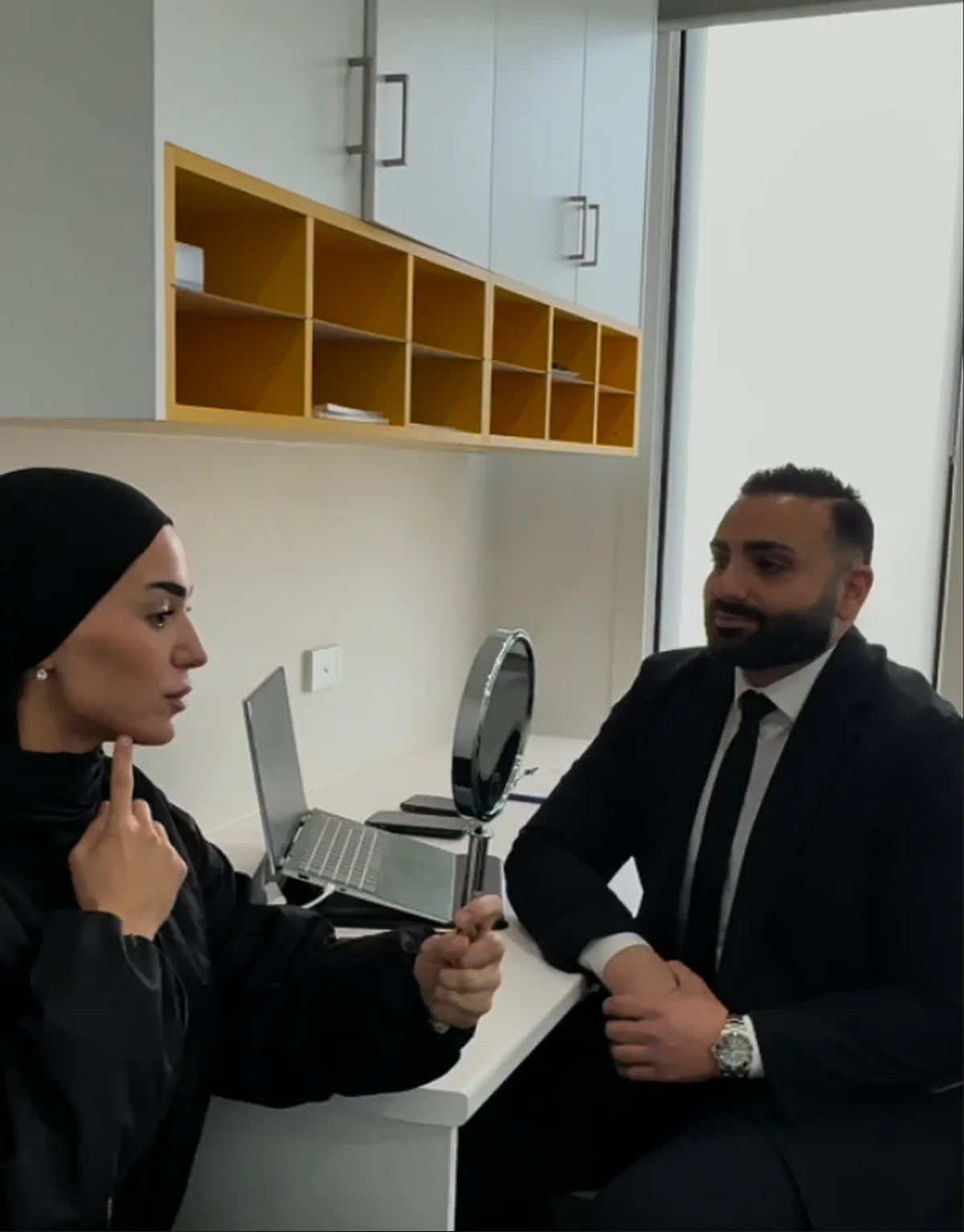
Patients call it several things: the nasolabial crease, nasal folds, naso folds, a naso labial fold, sometimes spelled nasallabial fold. They all describe the same structure.
Dr Ibrahim Hawli
MBBS  ·  Registered Medical Practitioner
tajmeel-nasolabial-folds
In short
A nasolabial fold is not a wrinkle. It is an anatomical structure made of dense fibrous tissue and muscle fibres that insert directly into the skin, and it is what allows the upper lip to rise when you smile. It is absent in newborns, disappears in a paralysed face, and remains after death. What changes with age is how deep it sits, driven by volume loss beside the nostril, bone recession beneath it, descent of the fat pad above it, and tension from the muscles that lift the upper lip.
01  /  Anatomy

What a nasolabial fold
actually is

Most people assume the fold is a line in the skin. It is not. A cadaver study examining full-thickness strips of facial tissue taken at right angles to the fold found it is made up of three things.
01

Dense fibrous tissue

A structural framework rather than a surface crease.
02

Levator muscle fibres

Fibres branching from the muscles that lift the upper lip.
03

Fold fascia bundles

Striated muscle bundles that originate in the fold’s own fascia.
Those muscle fibres do not stop at the muscle layer. The lip elevators and what the researchers called the fold muscles course down the lip, pass through the orbicularis oris, and insert directly into the dermis of the upper lip and into the border of the vermilion.
So the fold is a point where muscle attaches to skin. That is why it holds its shape, and why it behaves differently from a line creased into the surface over time.
02  /  The Key Point

Why the fold is normal anatomy,

not a wrinkle
The same study reported three findings that settle this question.
Finding one

Absent in a newborn

A baby has no nasolabial fold. It develops.
Finding two

Gone in paralysis

Where the facial nerve is not functioning, the fold flattens out. It depends on muscle activity to exist.
Finding three

Retained after death

A wrinkle caused by skin laxity would not be. A fibrous structure anchored to muscle is.
The study was titled the keystone of the smiling mechanism, and that is the point. The smile forms in stages, and the first stage is the levator muscles raising the upper lip as far as the fold. The fold is the hinge that movement happens around.
This matters clinically for one reason. A person asking for the fold to be removed entirely is asking for something that would interfere with how their face moves. Softening how deeply it sits is achievable. Erasing it is neither achievable nor desirable, and any practitioner promising it is describing a result nobody should want.
03  /  Causes

What causes a nasolabial
crease to deepen

Clinical literature separates this into distinct types, and the type present determines the approach.
Most patients have a combination. Establishing the proportion between them is the first job of an examination, and it is the difference between a plan that addresses the cause and one that addresses the appearance.
Type one

Volume deficiency beside the nostril

Reduced support immediately adjacent to the base of the nose. Patients with this pattern typically show limited superficial fat below the fold while the fat above it stays prominent, which creates a visible step.
Type two

Descent of the fat pad above the fold

The nasolabial fat compartment sits above the crease. When it loses support and moves downward, it stacks against the fold and deepens it from above. Skin laxity with age compounds this.
Type three

Tension from the upper lip elevators

Specific fibres of the levator labii superioris alaeque nasi, the levator labii superioris, and the zygomaticus major and minor exert continuous tension on the dermis every time the face moves. Because those fibres insert into the skin, that tension pulls on the fold directly.
04  /  Often Missed

Why the bone
underneath matters

This is the layer nobody mentions, and it explains a pattern that otherwise looks unexplainable.
The canine fossa is a shallow depression in the maxilla beneath the nostril. It provides the bony support for the soft tissue in that area. In some people that region recedes, and in others it is shallow from the outset.
Where the bone beneath the nostril offers less support, the soft tissue above it has nothing to sit on. The fold then reads as deep in a person who has not lost much fat at all, and who may be relatively young.
This is why an older photograph is useful at consultation, and why palpation is part of the examination rather than observation alone. A fold driven by bone shape is a different clinical situation from a fold driven by fat descent, even when they look similar in a mirror.
05  /  Not Uniform

Why different parts of nasal
folds behave differently

Repetitive muscle contraction produces lines running perpendicular to the direction of the muscle’s force. Because several muscles pass beneath and insert into different portions of the fold, contraction of a particular muscle affects a particular part of it.
Which muscle drives which portion
Medial portion
Levator labii superioris alaeque nasi
Middle portion
Levator labii superioris
Lateral portion
Zygomaticus muscles
One patient’s fold is deepest at the top near the nostril. Another’s is deepest at the bottom near the mouth. Those are different problems with different drivers, and treating the whole length as one unit ignores that.
At assessment you should be told which portion of your fold is most affected and which muscle is likely responsible.
06  /  Connected Areas

Why cheek volume
affects the fold

Many patients arrive concerned about the fold and are surprised when the cheek is examined.
A cadaver study of the deep medial cheek fat compartment established that when volume in that compartment reduces, the result is an excess skin envelope and the illusion of a more prominent fold beside the nose. The researchers named this pseudoptosis, meaning a false appearance of drooping.
In plain terms, the fold may not have changed at all. The support above and behind it has reduced, so the fold reads as deeper by comparison.
07  /  Why Care Matters Here

The vessels that run
alongside this fold

The facial artery courses along and in close proximity to the nasolabial fold, and its terminal branch, the angular artery, continues upward alongside the nose.
That is the reason this area is discussed in anatomical detail in clinical training rather than treated as a simple crease, and the reason a practitioner working here should be able to describe the vascular anatomy of the region without hesitation.
Documented in the literature

Why proximity to the facial artery matters

Clinical literature is direct about the consequence. Complications in this region arising from damage to the facial artery can lead to irreversible tissue loss or serious and permanent complications.
It is also worth understanding that the vessel position varies between individuals. Knowing where it usually runs is not the same as knowing where it runs in your face, which is why examination and palpation precede anything else.
08  /  Assessment

How the fold
is assessed

Grading scales exist for recording fold depth. They are useful for establishing a baseline and reviewing change over time rather than for deciding anything on their own.
09  /  Before You Decide

What to know before
your consultation

Every option carries potential risks as well as potential benefits, and no cosmetic medical procedure is risk free. In this region the relevant consideration is the vascular anatomy described above.
Bring a list of your current medications, and a photograph from several years ago if you have one.
Assessed at consultation

Factors that may make an approach inadvisable

That second-to-last point is not a formality. Complete removal is not an achievable or desirable outcome, and a consultation that begins with that expectation needs to address it before anything else.
Individual results vary. Not all people are suitable candidates. In some cases the appropriate recommendation is that nothing be undertaken.
10  /  What Happens

What happens at your consultation in Glenroy

Appointments run 30 to 45 minutes. A consultation fee applies and is confirmed when you book. No referral is required.
01

Discussion

What you have noticed, when it changed, any recent weight or health change, medical history, current medications and previous cosmetic work.
02

Assessment

The fold is examined at rest, in speech and in full smile. Portion depth, muscle contribution, support beneath the nostril, cheek behaviour, skin quality and asymmetry are assessed.
03

Information

What is clinically appropriate, what it involves, expected timeline, risks, alternatives and the option of proceeding with nothing.
04

Decision

You are not asked to decide on the day. Time to consider information is part of informed consent.
11  /  Service Area

Assessment for patients across Melbourne's northern suburbs

The clinic is on Glenroy Road, close to Glenroy Station and the Pascoe Vale Road corridor. Patients attend from Glenroy, Hadfield, Fawkner, Oak Park, Gowanbrae, Pascoe Vale, Coburg North, Broadmeadows, Campbellfield and Craigieburn.
12  /  Common Questions

Questions patients
ask

If your question is not answered here, ask it at your consultation. There is time set aside for exactly that.

No. It is an anatomical structure made of fibrous tissue and muscle fibres that insert into the skin. It is absent in newborns, flattens in a paralysed face, and remains after death. A wrinkle would not behave that way.

No, and it would not be desirable. The fold is part of how the upper lip rises when you smile. Softening how deeply it sits is achievable. Complete removal would interfere with facial movement.

Depth is not only about age. Shallow bony support beneath the nostril produces a deep fold in people who have lost very little volume, sometimes from a young age. Strong lip elevator muscles do the same. An older photograph usually clarifies which it is.

Asymmetry in this region is very common. Muscle activity is rarely identical on both sides, bone contour differs, and habitual sleeping position and chewing side contribute. It is often unnoticed until pointed out.

Yes. Research on the deep cheek fat compartment established that reduced volume there creates an excess skin envelope and the illusion of a more prominent fold. The fold may be unchanged. The support above it has reduced.

Muscle movement contributes, because the muscles that lift the upper lip insert directly into the skin of the fold and pull on it with every expression. But the fold exists because of that anatomy, not because of the smiling. People who smile less still have one.

Because the fold has at least four possible drivers, they look similar in a mirror, and distinguishing them requires palpation and observation in movement. The facial artery also runs close to this region and its position varies between individuals.

A consultation fee applies and is confirmed when you book.

Related concerns

The midface is assessed as a whole

Midface region
Lower perioral region
Perioral region
Temporal region
Dr Ibrahim Hawli
Written and clinically reviewed by
Dr Ibrahim Hawli, MBBS
Level 1 Certification, American Academy of Aesthetic Medicine. Advanced training in non surgical rhinoplasty with Aesthetic Intelligence.
Last reviewed: [insert month year]  ·  Next review due: [insert month year]

Your first step is

an assessment.

This page provides general information about cosmetic medical consultations and does not constitute medical advice. Any cosmetic medical procedure carries risks. Individual outcomes vary and not all people are suitable candidates. Any decision follows an individual consultation and assessment with a registered medical practitioner.